
Multimodal Deep Learning Model for Predicting Thrombus Thickness and Elasticity Using Ultrasound Imaging
English
Overview
This study proposes a multimodal deep learning model that predicts thrombus thickness and elasticity using ultrasound imaging.
The model integrates three distinct modalities — 1D LGFU signals, 2D frequency-domain spectra, and Doppler ultrasound data — to capture spatial, temporal, and flow-based information about blood clots.
By fusing these complementary features, the system achieves higher accuracy and robustness than single-modality baselines.
Model Architecture
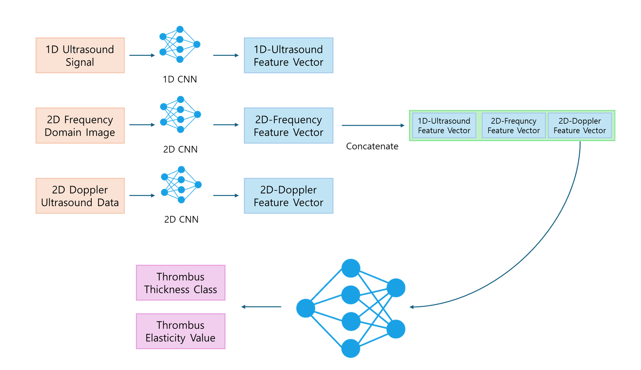
Figure 1. The proposed multimodal CNN architecture combining 1D, 2D, and Doppler ultrasound features.
1D LGFU Signal (Temporal Feature)
Laser-generated focused ultrasound (LGFU) signals capture reflected time-domain patterns corresponding to micro-level tissue changes.
Processed through a 1D CNN (4 convolutional layers, ReLU, Average Pooling).
2D Frequency-Domain Spectrum (Morphological Feature)
A Fast Fourier Transform (FFT) converts raw LGFU signals into 2D spectral images.
These are processed by a 2D CNN (3 conv layers, ReLU, MaxPool) to extract spatial-frequency structures.
Doppler Ultrasound (Flow Feature)
Doppler spectrograms obtained from Short-Time Fourier Transform (STFT) represent flow velocity and turbulence.
A specialized 2D CNN learns dynamic flow patterns linked to clot stiffness.
Fusion Layer
Feature vectors from all modalities are concatenated:
\[\mathbf{F_{fusion}} = \text{Concat}(F_{1D}, F_{2D_{FFT}}, F_{2D_{Doppler}})\]Outputs:
\[\hat{y}_{thickness} = \text{Softmax}(W_c \cdot \mathbf{F_{fusion}} + b_c), \quad \hat{y}_{elasticity} = W_r \cdot \mathbf{F_{fusion}} + b_r\]Pseudocode
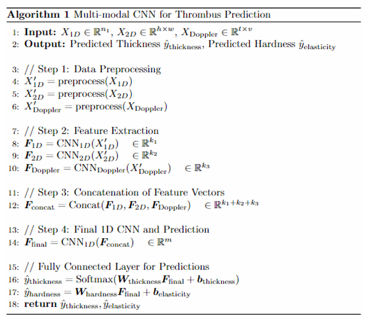
Figure 2. Pseudocode outlining data preprocessing, feature extraction, fusion, and final prediction.
Dataset and Simulation
- Data Source: Lab-generated artificial thrombi attached to vascular phantoms (100–1000 μm thickness).
- Modality Counts: 1D LGFU, 2D FFT spectra, Doppler spectrograms.
- Transforms: FFT for 1D → 2D conversion; STFT for Doppler.
- Training Details:
- 1D CNN (4 layers), 2D CNN (3 layers × 2 branches), Fully Connected Fusion.
- Optimizer: Adam (lr = $1 \times 10^{-4}$), BatchNorm + Dropout.
- Evaluation Metrics:
- Classification: Accuracy & F1-score for thrombus thickness.
- Regression: RMSE for elasticity prediction.
Results
| Model | Accuracy | F1-Score | RMSE (kPa) |
|---|---|---|---|
| 1D LGFU Only | 0.781 | 0.765 | 1.102 |
| 2D FFT Only | 0.826 | 0.801 | 0.972 |
| Doppler Only | 0.732 | 0.714 | 0.924 |
| Proposed Multimodal CNN | 0.853 | 0.828 | 0.854 |
The multimodal CNN achieved the best performance across both classification and regression tasks, confirming that integrating temporal, spatial, and hemodynamic modalities substantially improves predictive power.
Conclusion
This multimodal CNN framework provides a comprehensive understanding of thrombus properties by combining LGFU, FFT, and Doppler ultrasound data.
The integration of multiple modalities enables more precise and interpretable predictions of thrombus thickness and elasticity, offering potential applications in non-invasive cardiovascular diagnostics and AI-driven medical imaging.
Awards
🥈 KSEF 2025 Senior APB – Domestic Silver Medal
🥈 KSEF 2025 Senior APB – Inter Silver Medal
These distinctions recognize the study’s contribution to biomedical AI and its success in applying multimodal deep learning to real-world ultrasound data analysis.
한국어
연구 개요
본 연구는 초음파 영상 데이터를 이용해 혈전(Thrombus)의 두께와 탄성(Elasticity) 을 예측하는 다중모달 딥러닝 모델을 제안합니다.
모델은 1D LGFU 신호, 2D FFT 스펙트럼, 도플러 초음파 데이터를 결합하여,
시간적·공간적·혈류학적 정보를 모두 반영한 정밀한 혈전 분석을 수행합니다.
모델 구조

그림 1. 1D, 2D, Doppler CNN의 융합으로 구성된 다중모달 딥러닝 아키텍처
실험 및 결과
- 데이터: 인공 혈전 (100–1000 μm), LGFU 기반 초음파 측정
- 모델: 1D CNN + 2D CNN(FFT) + 2D CNN(Doppler) → Feature Fusion → Fully Connected Layer
- 지표: 정확도 (Accuracy), F1 점수, RMSE (kPa)
| 모델 | 정확도 | F1 점수 | RMSE (kPa) |
|---|---|---|---|
| 1D LGFU 단일 모델 | 0.781 | 0.765 | 1.102 |
| 2D FFT 단일 모델 | 0.826 | 0.801 | 0.972 |
| 도플러 단일 모델 | 0.732 | 0.714 | 0.924 |
| 제안된 다중모달 CNN | 0.853 | 0.828 | 0.854 |
결론
본 다중모달 CNN 모델은 LGFU, FFT, 도플러 초음파 데이터를 통합하여 혈전의 두께와 탄성을 정확히 예측할 수 있음을 보였습니다.
이는 향후 AI 기반 심혈관 질환 조기 진단 및 비침습 의료영상 분석 시스템으로의 확장 가능성을 제시합니다.
수상 내역
🥈 2025년 KSEF Senior APB 국내 부문 은상
🥈 2025년 KSEF Senior APB 국제 부문 은상
본 연구는 다중모달 딥러닝과 의료영상 융합의 혁신성으로 높은 평가를 받았습니다.